The Royal Microscopical Society (RMS), based in Oxford, is a charity dedicated to furthering the science of microscopy. They achieve this through a wide range of activities supporting research and education in microscopy, and through enabling microscopists to make advances and developments in microscopy, cytometry, and imaging.
Here, the RMS have provided us with a peep through the lens, can you guess what you’re looking at?
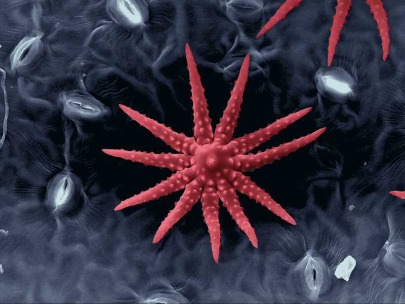
Credit – Stephanie Schuller
Answer – These are epidermal hairs on the underside of a plant leaf. They help to limit prey by insects and damage by parasites.
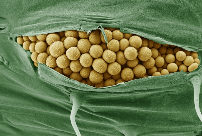
Credit – Kim Findlay
Answer – Cryo-SEM of yellow rust infection on wheat. Taken on a Zeiss Supra 55 FEG SEM with a Gatan Alto 2500 cryo-system. Prepared by plunge freezing in liquid nitrogen slush (coloured using Photoshop).
![]()
Credit – Benjamin Myers
Answer –This is an example of single crystal silicon after etching with potassium hydroxide (KOH). Small KOH crystals can be seen on the surface after drying. The image was captured with a scanning electron microscope (SEM) which is used to inspect etch quality. This image was constructed by acquiring images with multiple detectors and overlaying to achieve false colour.
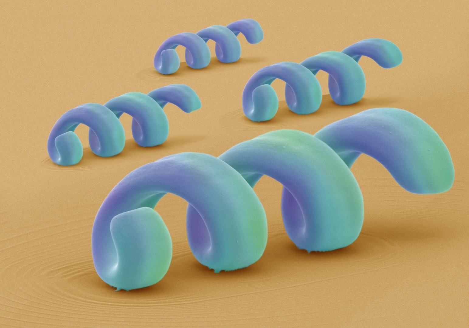
Credit – David McCarthy
Answer – This image is of ‘Nano Swimmers’ which are currently being investigated for potential use as novel drug carriers. These coiled structures are 25 microns in length, 5 microns in diameter and 300 nanometres in thickness. They are composed of a polymer with nickel/titanium coating and were fabricated by the Multi-Scale Robotics Laboratory, ETH Zurich and in collaboration with the NanoMedicine Laboratory, UCL School of Pharmacy. The swimmers were imaged after each sample was given a 5nm gold coating. In addition, a tilt angle of 65o enhanced their full structure. The images were imported into photoshop, where they were artistically manipulated and coloured by Annie Cavanagh.
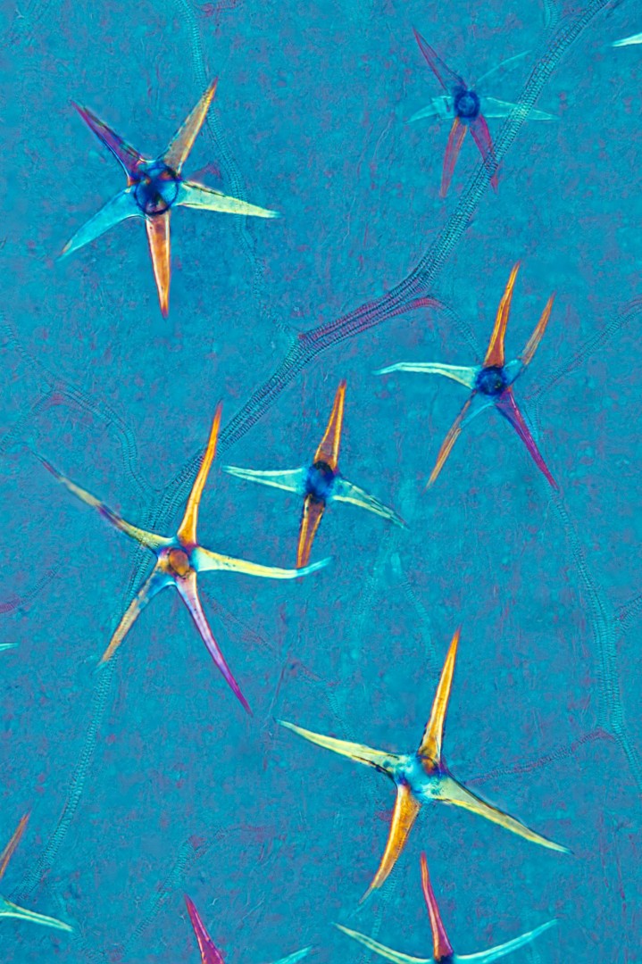
Credit – Steve Lowry
Answer – Leaf of Deutzia scaber showing leaf scales. This polarised light micrograph uses wave retarding filter. The stacked image is comprised of five exposures.
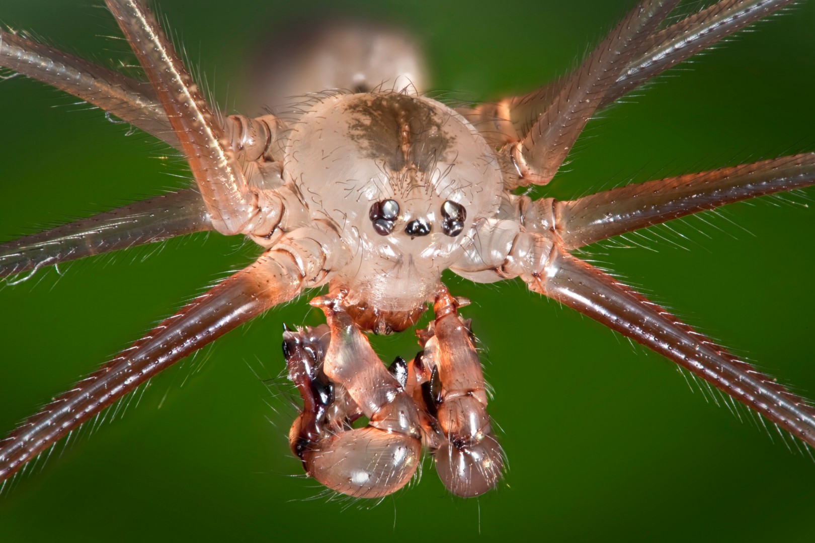
Credit – Harold Taylor
Answer – Daddy-long-legs Spider, also known as the Pholcus phalangioides. Wild M420, Z stack
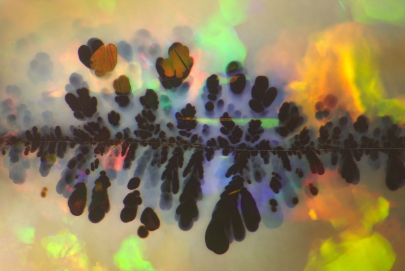
Credit – Nathan Renfro
Answer – This black manganese oxide was carried by an aqueous solution through a crack in an Australian precious opal to show the iridescent effect known as "play of colour". The manganese oxide penetrated into the opal before solidification leaving black manganese oxide plumes along the crack that introduced the foreign metal oxide. Field of View: 5.85mm
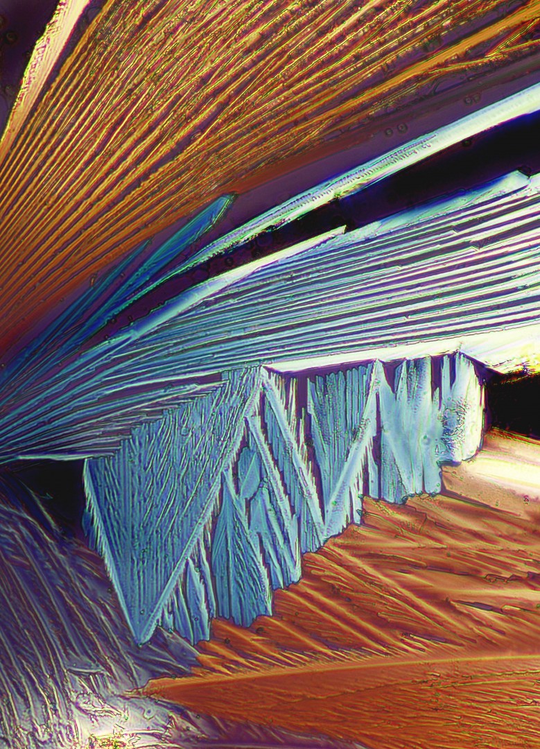
Credit – Michael Reese Much FRMS
Answer – Elon crystals photographed using cross-polarization microscopy. Elon is a primary component of many black-and-white photographic developers.
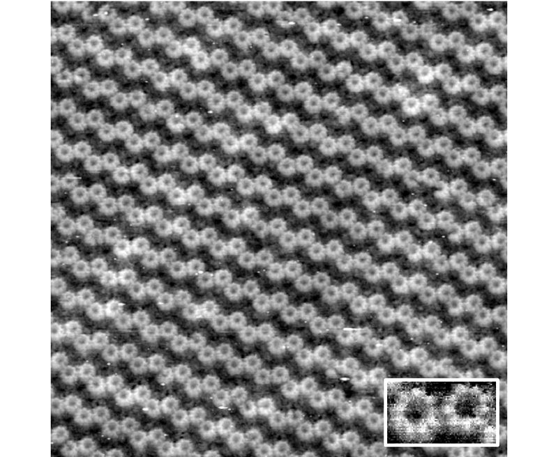
Credit – Sandip Kumar
Answer – Highest resolution image of single LH2 nonamer rings where defects in the ring are visible and single subunits can be discerned.
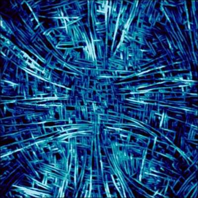
Credit – Nic Mullin
Answer – Atomic force microscope height image of the core of a type I spherulite in a thin film of isotactic polypropylene. The bright features are lamella crystals lying edge-on. The cross-hatching is due to homoepitaxy on the (010) crystal plane of the iPP lamella crystals. Lateral scale 3.5μm, colour scale 18nm.
More about The RMS…
The RMS is a not-for-profit organisation and is a registered Charity (number 241990~) under the Charities Act 2011. The Society provides a great community for microscope users, with a busy calendar of training courses and networking events each year. The RMS also produce the Journal of Microscopy, infocus magazine exclusively for their members and a wide range of microscopy handbooks.
For more information, visit rms.org.uk


